Precystic form considered transient, it develops after the luminal. Amoebae are distinguished by their minimum size, no more than 10-18 microns. It is difficult to detect them, due to their insignificant content in the feces.
Existing varieties of protozoa
- amoeba proteus;
- dysentery;
- intestinal.
Amoeba proteus

Dysentery amoeba
Dysentery amoeba
Dominates exclusively in the human large intestine and water bodies. Once in the body, it causes a serious disease of amoebiasis. In its life cycle, three main stages are recorded: cyst, small vegetative and large vegetative forms, tissue.
Penetration into the body is carried out through the consumption of contaminated food in the form of cysts. In terms of its dimensions, it is characterized by minimal dimensions. The small vegetative form does not cause negative symptoms from the body; it settles in the lower intestine.
Intestinal amoeba
Non-pathogenic amoeba
There are certain types of amoebas that belong to the non-pathogenic class. This category includes:
Amoeba Hartmann
With a detailed study, experts are able to make the wrong diagnosis. This is due to the lack of specific external data.

Common amoeba
Dwarf amoeba
Diagnosis is made by using Lugol's solution. A distinctive feature of the amoeba is its small size and the presence of a distinct shell.
Iodameba Bütschli
Dientameba
When released into the environment, bacteria die or are destroyed, they are not adapted to adverse conditions.
Oral amoeba
It occurs in almost all people who suffer from diseases of the oral cavity. In some cases, the bacterium is found in lesions of the respiratory system. Its size does not exceed 30 microns, the nuclei are practically invisible, the movement is slow.
Penetrating into the human body, bacteria lead to serious disruptions in the functioning of the digestive system. The most common type of disease is. It comes in several varieties:
Acute form
The acute form of the disease begins spontaneously. At first, a person is constantly bothered by a violation of the stool with predominant diarrhea. Gradually, pain syndrome is added to the general clinical picture. The feces contain a small amount of blood and mucus. If the disease develops in children, fever and vomiting are observed.
Lightning shape
The fulminant form is characterized by a severe course. It is characterized by the presence of acute toxic syndrome, with serious damage to the intestinal walls. Are prone to the development of pathology in women in the postpartum period.
In the absence of a therapeutic effect, a high risk of death remains.
Lingering amebiasis
Prolonged amebiasis is accompanied by severe intestinal motility disorders. The person often has constipation and diarrhea. In this case, acute pain syndrome, nausea and weakness are recorded. The patient refuses to eat.

Extraintestinal amebiasis is characterized by damage to many organs, in particular the liver
Extraintestinal amebiasis
A less common type of disease is extraintestinal amebiasis. It is characterized by damage to many organs, in particular the liver. Severe violations are recorded exclusively in adults, and require immediate surgical intervention.
Coping with amoebas is not so easy, due to their high resistance to adverse conditions.
In contact with
Amoebas are a genus of unicellular eukaryotic organisms (classified as protozoa). They are considered animal-like, as they feed heterotrophically.
The structure of amoebas is usually considered on the example of a typical representative - an ordinary amoeba (proteus amoeba).
Common amoeba (hereinafter referred to as amoeba) lives at the bottom of freshwater reservoirs with contaminated water. Its size ranges from 0.2 mm to 0.5 mm. In appearance, the amoeba looks like a shapeless, colorless lump that can change its shape.
The amoeba cell does not have a rigid shell. It forms protrusions and protrusions. Bulges (cytoplasmic outgrowths) are called pseudopods or pseudopodia... Thanks to them, the amoeba can slowly move, as if flowing from place to place, and also grab food. The formation of pseudopods and the movement of the amoeba occurs due to the movement of the cytoplasm, which gradually flows into the protrusion.
Although the amoeba is a unicellular organism, and there can be no question of organs and their systems, almost all vital processes characteristic of multicellular animals are characteristic of it. Amoeba eats, breathes, secretes substances, multiplies.
The cytoplasm of the amoeba is not homogeneous. A more transparent and dense outer layer is distinguished ( eqtoplasma) and a more granular and liquid inner layer of the cytoplasm ( endoplasm).
The cytoplasm of the amoeba contains various organelles, the nucleus, as well as the digestive and contractile vacuoles.
The amoeba feeds on various unicellular organisms and organic debris. Food is clasped by pseudopods and is inside the cell, it is formed digestiveand I vacuole... It receives various enzymes that break down nutrients. Those that are needed by the amoeba then enter the cytoplasm. Unnecessary food debris remains in the vacuole, which comes to the cell surface and everything is thrown out of it.
The "organ" of excretion in amoeba is contractile vacuole... Excess water, unnecessary and harmful substances from the cytoplasm enter it. The filled contractile vacuole periodically approaches the cytoplasmic membrane of the amoeba and pushes out its contents.

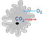
The amoeba breathes with the entire surface of the body. Oxygen enters it from the water, and carbon dioxide from it. The respiration process consists in the oxidation of organic substances in mitochondria with oxygen. As a result, energy is released, which is stored in ATP, and also water and carbon dioxide are formed. The energy stored in ATP is then spent on various vital processes.
For the amoeba, only asexual reproduction is described by dividing in two. Only large, i.e. grown, individuals are divided. First, the nucleus is divided, after which the amoeba cell is divided by a constriction. The daughter cell that does not receive a contractile vacuole forms it later.
With the onset of cold weather or drought, the amoeba forms cyst... Cysts have a dense membrane that performs a protective function. They are quite lightweight and can be carried by the wind over long distances.
Amoeba is able to react to light (crawls away from it), mechanical irritation, the presence of certain substances in water.
abstract
On the topic: Amoeba
Completed by: 1st year student A.R.Davletkulova
Checked by: V.N. Satarov
Ufa-2012
2.structure and vital functions of the amoeba
3.Dysentiric amoeba
Amoeba
In addition to pseudopodia, because of which the body of the amoeba does not have a definite shape, these organisms are characterized by the absence of a rigid cell membrane. The cell is surrounded only by a special molecular layer, the plasma membrane - an integral part of the living cytoplasm. The latter is subdivided into a thin surface, relatively homogeneous part, called ectoplasm, and a granular endoplasm lying in the depths. That, in turn, consists of an outer gelatinous zone, a plasmagel, and an inner fluid plasmazole. The endoplasm contains the nucleus, as well as the digestive and contractile vacuoles. Food captured by pseudopodia, such as bacteria, algae and protozoa, is surrounded by a digestive vacuole and is digested in it. Undigested material is ejected from the cell when the membrane of this vacuole fuses with the plasma membrane. Metabolic waste is excreted by simple diffusion. A certain part of them, possibly, is removed through the contractile vacuoles, but the main function of the latter is to remove excess water from the cell. They contract from time to time, pushing it out. Reproduction in amoebas is asexual - by dividing the cell in two. At the same time, the nucleus is divided mitotically, and then the cytoplasm is pulled over and splits into two parts that are approximately the same in volume, each containing a daughter nucleus. The two cells that are formed grow and eventually divide too.
The structure and life of the amoeba
It is a gelatinous, single-celled creature, so small that it can only be seen under a microscope. The main species of amoeba live in freshwater rivers and ponds. But there are species that live at the bottom of salt water bodies, in moist soil and food. Amoeba is constantly changing its shape. She moves, pushing forward first one of her half, then the other. Like many jelly-like organisms, the amoeba moves in such a way that it forms a shape called a "false stalk," or pseudopodia. When the pseudopod reaches food, it envelops it and takes it into the main body. This is how the amoeba feeds. She has no mouth. Amoeba belongs to the class of protozoa, which are the lowest class of living beings. It has no lungs or gills. But it absorbs oxygen from water, releases carbon dioxide, digests food, as more complex animals do. The amoeba probably also has feelings. When touched or aroused, she immediately curls up into a tiny ball. Amoeba avoids bright light, too hot or cold water. In an adult amoeba, the nucleus, a tiny dot in the center of the protoplasm, divides in two. After that, the amoeba itself bifurcates, forming new independent organisms. When they reach their full size, they start dividing again. In their structure, protozoa are extremely diverse. The smallest ones have a diameter of 2-4 microns (a micrometer is 0.001 mm). Their most common sizes are in the range of 50-150 microns, some reach 1.5 mm and are visible with the naked eye.
The amoeba has the simplest structure. The body of the amoeba is a lump of semi-liquid cytoplasm with a nucleus in the middle. The entire cytoplasm is subdivided into two layers: the outer, viscous - ectoplasm and the inner, much more liquid - endoplasm. These two layers are not sharply demarcated and can transform into each other. The amoeba does not have a hard shell, and it is able to change the shape of the body. When an amoeba crawls on a leaf of an aquatic plant, bulges of cytoplasm are formed in the direction in which it moves. Gradually, the rest of the amoeba cytoplasm flows into them. Such protrusions are called pseudopods or pseudopodia. With the help of pseudopodia, the amoeba not only moves, but also captures food. With pseudopodia, it covers a bacterium or microscopic alga, soon the prey is inside the body of the amoeba, and a bubble forms around it - a digestive vacuole. Undigested food residues are thrown out after a while.
Amoeba proteus: 1 - core; 2 - digestive vacuoles; 3 - contractile vacuole; 4 - pseudopods; 5 - undigested food debris thrown out.
In the cytoplasm of the amoeba, a light bubble is usually visible, which appears and disappears. This is a contractile vacuole. It collects excess water that accumulates in the body, as well as liquid waste products of the amoeba. Amoeba breathes, like all other protozoa, the entire surface of the body.
Euglena green: 1 - flagellum; 2 - an eye spot; 3 - contractile vacuole;
The most complex structure of the protozoa in ciliates. Unlike the amoeba, their body is covered with the thinnest shell and has a more or less constant shape. Support and shape of the body is also supported by supporting fibers running in different directions. However, the body of ciliates can quickly contract, change its shape, and then return to its original shape. Contraction is carried out with the help of special fibers, similar in many respects to the muscles of multicellular animals. Ciliates can move very quickly. So, a shoe in a second overcomes a distance exceeding the length of its body by 10-15 times. At the same time, a lot of cilia that cover the entire body of the ciliate perform fast rowing movements, up to 30 per second (at room temperature). In the ectoplasm of the shoe, there are many trichocyst rods. When irritated, they are thrown outward, turning into long threads, and hit the enemy who attacks the ciliates. Instead of those thrown out in the ectoplasm, new trichocysts are formed. On one side, approximately in the middle of the body, the shoe has a deep oral cavity leading into a small tubular pharynx.
Infusoria shoe: 1 - cilia; 2 - digestive vacuoles; 3 - large nucleus (macronucleus); (micronucleus); 5 - mouth opening and pharynx; 6 - undigested food debris thrown out; 7 - trichocysts; 8 - contractile vacuole.
Through the pharynx, food enters the endoplasm, where it is digested in the formed digestive vacuole. In ciliates, unlike amoebas, undigested food debris is thrown away at a certain place in the body. Their contractile vacuole is more complex and consists of a central reservoir and conducting channels. The ciliate has two types of nuclei: large - macronucleus and small - micronucleus. Some ciliates may have several macro- and micronuclei. The macronucleus differs from the micronucleus in a significantly larger number of chromosomes. And therefore, it contains a lot of deoxyribonucleic acid (DNA), which is part of the chromosomes.
Various types of ciliates: 1 - ciliate trumpeter; 2-5 - planktonic ciliates.
Dysenteric amoeba (Entamoeba histolytica), the simplest of the order of amoeba; the causative agent of amoebic dysentery was first described in 1875 by the Russian scientist F.A. Leshem. When it enters the intestine of a person D. and. in most cases, it multiplies in the contents of the large intestine, without penetrating into tissues and without causing disturbances in intestinal function (a person is healthy, but serves as a carrier of D. and.) This form D. and. called luminal (forma minuta) (size about 20 microns) (Fig. 1, a). It moves with the help of pseudopodia. The nucleus is spherical, 3-5 µm in diameter; chromatin is located under the nuclear envelope in the form of small lumps; in the center of the nucleus there is a small karyosome. There may be several phagocytosed bacteria in the endoplasm. When faeces thicken in the colon, the luminal form is surrounded by a membrane and turns into a spherical cyst (size about 12 microns) with 4 nuclei that do not differ in structure from the nucleus of the vegetative form; immature cysts contain 1-2 or 3 nuclei. There is a vacuole with glycogen; some of the cysts contain short, bar-shaped formations - chromatoid bodies (Fig. 1, b). With feces, cysts are thrown out into the external environment and can again enter the human gastrointestinal tract, where, after the metacystic stage of development (division into 8 daughter amoebas), they give rise to luminal forms (Fig. 2, A).
Dysentery amoeba was first described by the Russian scientist L.F. Lesh (1875).
The structure of the dysentery amoeba.
The amoeba comes in various forms.
Large vegetative form of amoeba .
The large vegetative form of the amoeba is larger, its size is 20-60 microns. The cytoplasm of the amoeba is divided into 2 layers: external and internal. The amoeba is transparent, colorless; the nucleus of a living amoeba is not visible. In a dead and motionless amoeba, the nucleus appears in the form of a ring-shaped cluster of shiny grains. The endoplasm often contains from one to several red blood cells at different stages of digestion, which is very typical of the amoeba form. This amoeba is often called a hematophage, or erythrophage (erythrocyte eater). It differs from other types of amoeba in forward motion. Under the microscope, one can see how the ectoplasm outgrowth is jerkingly formed and the entire endoplasm is poured into it quickly with a swirl. Then a new pseudopod is formed, and again the contents of the amoeba are quickly transfused. Sometimes the amoeba seems to freeze for a few moments, and then suddenly begins its characteristic movement again. A large vegetative form is found in freshly excreted liquid feces of a patient with acute amebiasis, which undoubtedly confirms the diagnosis.
Tissue form of amoeba.
 Translucent form of amoeba.
The lumen form of amoeba lives in the lumen of the upper sections of the large intestine and is the main form of existence of dysentery amoeba. Luminal forms can be found in the liquid freshly excreted feces of convalescents or patients with chronic amoebic dysentery. In carriers or patients in remission, it does not occur in formalized or semi-formed stools. For detection, it is necessary to examine feces obtained by deep lavage of the intestines, or the last portions of feces after taking a saline laxative. The size of the amoeba is 15 - 20 microns. In the native preparation, the amoeba nucleus is not visible. The cytoplasm contains bacteria, small vacuoles, but does not contain erythrocytes. The movement is weaker than that of the tissue form, pseudopods are formed more slowly, their size is also smaller. The division into ecto- and endoplasm is expressed only with the formation of pseudopods.
Precystic form amoeba .
The precystic form of amoeba is usually found in semi-formed stools. The size of the amoeba is 12 - 20 microns. In structure, it resembles a luminal form.
The life cycle of a dysentery amoeba.
The luminal forms of dysentery amoeba live in the upper part of the large intestine of a person, without causing him harm. However, under some conditions, turning into pathogenic tissue forms, they penetrate the intestinal wall.
The luminal forms, passively moving along with the contents of the intestine, enter its end sections, where unfavorable conditions (dehydration, changes in the bacterial flora, changes in the pH of the environment, etc.) lead to the death of amoebas or their transformation into cysts. Cysts with human feces are released into the environment, where they can persist for a long time. Mature quadruple cysts are infectious to humans.
Cysts, getting into water, on vegetables, hands and food (to which they are carried, in particular, by flies), various objects, for example dishes, toys, are eventually brought into the human mouth. From here they enter the gastrointestinal tract, where their membrane dissolves. Each nucleus is divided in two, an eight-core amoeba is formed, from which 8 daughter ones arise.
Clinical picture dysentery amoeba.
Dysentery, or histological, amoeba causes a disease in humans, amoebic dysentery, or amebiasis. Multiple ulcers form in the large intestine. The disease is of varying severity and begins acutely or gradually. Disturbed by pain in the lower abdomen, frequent liquid stools of a red-brown color due to the admixture of blood and mucus (while feces often resemble meat slops). Body temperature is usually normal. The disease can last with periodic exacerbations for several years. In severe cases, anemia and exhaustion develop.
The tissue form of amoeba from intestinal ulcers can be carried with blood into the liver, lungs, brain and other organs, causing abscesses there. These complications can be fatal without timely treatment.
Diagnosis.
To identify dysentery amoebas or their cysts, excrement is examined. For this purpose, native smears of feces are prepared on glass slides in a drop of isotonic sodium chloride solution and a drop of Lugol's solution.
In the native smear (X400), mobile vegetative forms are observed. Cysts are clearly visible in Lugol's solution. In difficult cases, the preparations are stained according to Heidenhain.
For research, you need to take freshly excreted feces, since amoeba quickly, within 10-20 minutes, lose mobility, which makes reliable diagnosis impossible. Cysts of amoebas can also be found in formalized feces, even if stored for several hours before examination. If only luminal forms or cysts are identified, then it is impossible to diagnose amoebic dysentery, since they can only be a sign of carriage. Therefore, with clinical indications, that is, a suspicion of the possibility of amoebiasis, repeated studies are carried out, a saline laxative is prescribed, because large vegetative or tissue forms can be found only in liquid or semi-liquid feces. In this case, first of all, pathological impurities (lumps of mucus) are examined.
It should be borne in mind that in the acute stage of the disease, only tissue, or rather, large vegetative forms, are more often excreted with feces, and during the recovery period - luminal forms and cysts.
If it is impossible to immediately study the feces, their conservation is allowed. Canned material can be examined after a few days or sent for consultation. The simplest in the preservative are colored and lose their mobility, which, to a certain extent, complicates laboratory research.
If an amoebic abscess is suspected, the contents obtained during surgery or puncture are microscoped. At the same time, amoebas are more often found in the material taken at the border of healthy and affected tissues, on the inner surface of the abscess capsule, than directly into the pus. Previous antibiotic or chemotherapy may lead to a negative result of such a study. Methods for serological diagnosis of amoebiasis (RHA, RIF, REMA) have been developed.
Prevention.
The distribution and mechanism of transmission of amoebic and bacterial dysentery have much in common, therefore preventive measures are also similar. The patients are hospitalized. Discharge is allowed after receiving 3 negative feces tests carried out within 1 week. In case of unstable stool in convalescents, as well as if it is necessary to identify carriers among healthy individuals, at least 6 tests are performed within 2 weeks.
After discharge, those who have recovered are subject to observation in the offices of infectious diseases of polyclinics for at least a year with periodic examination of feces. The carriers are sanitized.
Feces, contaminated linen are neutralized with a 3% lysol solution. The usual chlorination of water does not affect cysts. Only boiling gives a quick effect.
Carriage of dysentery amoeba is recorded everywhere, but diseases are observed most often in Central Asia, the Caucasus and the Far East. Imported cases are possible.
The sub-kingdom of Unicellulars includes animals whose body consists of only one cell, mostly microscopic in size, but with all the functions inherent in the body. Physiologically, this cell is a whole independent organism.
The two main body components of unicellular organisms are the cytoplasm and the nucleus (one or more). The cytoplasm is surrounded by an outer membrane. It has two layers: the outer (lighter and denser) - ectoplasm - and the inner - endoplasm. The endoplasm contains cellular organelles: mitochondria, endoplasmic reticulum, ribosomes, elements of the Golgi apparatus, various supporting and contractile fibers, contractile and digestive vacuoles, etc.
Habitat and external structure of the common amoeba
The simplest lives in water. It can be lake water, a dew drop, soil moisture, and even water inside us. Their body surface is very delicate and dries instantly without water. Outwardly, the amoeba looks like a grayish gelatinous lump (0.2-05 mm), which does not have a permanent shape.
Motion
The amoeba "flows" along the bottom. On the body, outgrowths that change their shape are constantly formed - pseudopodia (pseudopods). Cytoplasm is gradually poured into one of these protrusions, the false pedicle attaches to the substrate at several points and movement occurs.
Internal structure
The internal structure of the amoeba
Food
Moving, the amoeba encounters unicellular algae, bacteria, small unicellular organisms, "flows around" them and includes them in the cytoplasm, forming a digestive vacuole.

Amoeba food
Enzymes that break down proteins, carbohydrates and lipids enter the digestive vacuole, and intracellular digestion occurs. Food is digested and absorbed into the cytoplasm. The method of capturing food using false legs is called phagocytosis.
Breath
Oxygen is consumed in cellular respiration. When it becomes less than in the external environment, new molecules pass into the cell.
Amoeba breath
The molecules of carbon dioxide and harmful substances accumulated as a result of vital activity, on the contrary, go out.
Highlighting
The digestive vacuole approaches the cell membrane and opens outward so that undigested residues are thrown out in any part of the body. The fluid enters the body of the amoeba through the formed thin tubular canals, through pinocytosis. The contractile vacuoles are involved in pumping out excess water from the body. They gradually fill up, and every 5-10 minutes they sharply contract and push the water out. Vacuoles can occur anywhere in the cell.
Reproduction
Amoebas reproduce only asexually.

Reproduction of amoeba
The grown amoeba begins to reproduce. It occurs through cell division. Before cell division, the nucleus doubles so that each daughter cell receives its own copy of hereditary information (1). Reproduction begins with a change in the kernel. It stretches out (2), and then gradually lengthens (3,4) and tightens in the middle. The transverse groove is divided into two halves, which diverge in different directions - two new nuclei are formed. The body of the amoeba is divided into two parts by the constriction and two new amoeba are formed. Each of them contains one core (5). During division, the formation of missing organelles occurs.
The division can be repeated several times during the day.
Asexual reproduction - a simple and quick way to increase the number of your descendants. This method of reproduction does not differ from cell division during the growth of the body of a multicellular organism. The difference is that the daughter cells of a single-celled organism diverge as independent cells.
Reaction to irritation
Amoeba has irritability - the ability to feel and respond to signals from the external environment. Crawling on objects, it distinguishes edible from inedible and captures them with pseudopods. She crawls away and hides from the bright light (1)
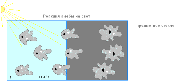
mechanical irritation and increased concentration of substances harmful to it (2).
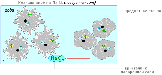
This behavior, which consists in moving towards or away from the stimulus, is called taxis.
Sexual process
Absent.
Experiencing adverse conditions
A single-celled animal is highly sensitive to environmental changes.
In unfavorable conditions (when the reservoir dries up, in the cold season), amoebas draw in pseudopodia. A significant amount of water and substances are released from the cytoplasm onto the body surface, which form a strong double shell. There is a transition to a resting state - a cyst (1). In a cyst, life processes are suspended.
Cysts carried by the wind contribute to the settlement of the amoeba.

When favorable conditions occur, the amoeba leaves the cyst membrane. It releases pseudopodia and goes into an active state (2-3).
Another form of protection is the ability to regenerate (recover). A damaged cell can complete its destroyed part, but only if the nucleus is preserved, since all information about the structure is stored there.
Life cycle of amoeba
The life cycle of an amoeba is simple. The cell grows, develops (1) and divides asexually (2). In bad conditions, any organism can "temporarily die" - turn into a cyst (3). When conditions improve, it "comes back to life" and multiplies vigorously.