Experienced doctors know all kinds of hypoxia. This pathological condition underlies the development of many diseases. Hypoxia is the reduction of oxygen in the tissues. All this is reflected in the state of vital organs (brain, spinal cord, heart, kidneys).
What types of hypoxia are
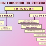
There are several types of this disease. Hypoxia is endogenous (due to internal factors) and exogenous. Endogenous hypoxia is divided into the following types:
- respiratory;
- cardiac (circulatory);
- blood (hemic);
- load;
- tissue;
- substrate;
- mixed
A type of exogenous hypoxia is man-made. The rate of development of this state is different. Depending on this, the following types of oxygen starvation are distinguished:
- instantaneous;
- spicy;
- subacute
- chronic.
Instant fasting is characterized by rapid development (within 1-3 minutes). The duration of the acute form of hypoxia is less than 2 hours. Subacute form of oxygen deficiency develops within 3-5 hours. Chronic can occur for months and even years. This pathology proceeds in 2 stages.
The first stage is characterized by increased respiration and heartbeat, as well as redistribution of blood flow. In response to a lack of oxygen, more red blood cells and hemoglobin are formed, which contributes to an increase in the oxygen capacity of the blood. This is a protective reaction of the body. At the stage of decompensation, various organs are affected.
There are also hypoxia mild, moderate, severe and critical degree. In the first case, signs of lack of oxygen occur during exercise. A critical degree of hypoxia can cause shock or coma. Death is possible.
Exogenous form of hypoxia
The causes of this pathology are different. Very often hypoxic hypoxia develops. Otherwise, it is called external. This condition is due to the low concentration of oxygen in the ambient air. When breathing, the blood is not properly saturated with oxygen. There are 3 types of exogenous hypoxia:
- hyperbaric;
- normobaric;
- hypobaric.
The basis of this separation is the level of oxygen pressure. It can remain normal if a person is in a stuffy, confined space (mines, wells). A decrease in pO2 is observed when climbing to a height. The higher a person’s above sea level, the less oxygen is in the air.
Climbers, pilots, parachutists, mountain dwellers, tourists often face this problem. In this case, hypoxia can cause mountain (altitude) disease. Acute oxygen starvation can occur when flying on aircraft without special oxygen masks. Less common are situations when oxygen pressure is elevated.
This is possible when using medical equipment. A breakdown or malfunction of the apparatus during hyperbaric oxygenation leads to an excess of oxygen, which has a toxic effect on the body. Persons living in megalopolises and near industrial enterprises often encounter chronic exogenous hypoxia. Oxygen starvationdue to exogenous factors, manifested by the following features:
- dizziness;
- headache;
- loss of consciousness;
- blue skin.
Residents of highlands develop adaptive reactions. This ensures the adaptation of the body to hypoxia.
What is different circulatory hypoxia
Many diseases of the cardiovascular system are accompanied by hypoxia. The following causes of lack of oxygen are distinguished:
- decrease in the volume of the circulating blood;
- increased blood viscosity;
- severe dehydration;
- the presence of blood clots;
- venous congestion.
Oxygen deficiency is local and widespread. It all depends on the degree of circulatory disorders. Only 1 organ can suffer. The basis of the development of this form of hypoxia are the following pathological processes: congestion and ischemia. Ischemic type of hypoxia develops when the volume of blood entering the organ decreases.
The cause may be heart attack, heart sclerosis, left ventricular failure, shock, a sharp drop in blood pressure, vasoconstriction. At the same time, blood oxygen saturation is normal. Congestive hypoxia is a consequence of a decrease in blood flow velocity.This is possible with failure of the right ventricle, thrombophlebitis of the lower extremities.
The development of tissue hypoxia
The tissue form of oxygen starvation of tissues is caused by the breakdown of oxidative processes. At the same time, tissues and organs are supplied with oxygen in sufficient quantity. The basis of the development of this pathology is a decrease in the activity of enzymes. There are several causes of tissue hypoxia. There are the following etiological factors in the development of this pathology:
- lack of vitamins in the body (thiamine, riboflavin, nicotinic acid);
- cyanide, alcohol, ether, or urethane poisoning;
- drug poisoning (barbiturates);
- high dose drug use;
- exposure to bacterial toxins;
- thyrotoxicosis;
- radiation exposure;
- severe infectious diseases;
- poisoning of the body with protein metabolism products;
- extreme depletion of the body (cachexia).
Tissue hypoxia can develop gradually over a long time.
Other forms of hypoxia
Often develops hemic type of oxygen starvation of tissues. If tissue hypoxia is caused by a decrease in the activity of the enzyme system, then in this situation the reason lies in the change in the composition of the blood itself. The main reason is a decrease in hemoglobin concentration. This is possible against the background of anemia. Blood hemoglobin is known to carry oxygen. Its lack leads to a decrease in tissue oxygen saturation.
Reducing the level of beneficial hemoglobin is possible with carbon monoxide poisoning, nitrates, sulfur.
Predisposing factors include smoking, inhalation of exhaust gases, inhalation of smoke during a fire. Drug poisoning can cause hypoxia. Sometimes substrate hypoxia develops. When it receives a sufficient amount of oxygen, but there is a shortage of nutrients that must be oxidized by oxygen.
This video describes hypoxia and its consequences:
This is possible with diabetes, prolonged fasting or a strict diet. Often develop hypoxia load. It is associated with an increase in cell oxygen demand. Often this is observed on the background of thyrotoxicosis or with heavy physical labor. A lack of oxygen can be experienced by the fetus during pregnancy. Thus, oxygen starvation of tissues is a sign of the most diverse pathologies.
Hemic hypoxia is one of the types of hypoxia, which is primarily a pathological process characterized by oxygen starvation of certain cells and organ systems of the body. The causes of this condition can be quite diverse and will depend on the type of hypoxia and the system of the body on which it affects.
Types of the pathological process
At the moment there is not one classification of this pathological process, but most often they use the one created on the basis of the mechanisms and causes of development. Based on this classification, there are such types as:
- Hypoxia exogenous. It includes two types: it is hypoxic and hyperoxic. This type of oxygen starvation develops when the oxygen content in the air inhaled by the body changes under the influence of external conditions and when the barometric pressure changes.
- Respiratory hypoxia, which is called respiratory. It develops when a violation of the penetration of oxygen into the blood through the respiratory tract of a person occurs. This can occur due to a violation of the structure of the respiratory apparatus, its pathology, for example, with partial obstruction of the paths with a tumor, any foreign bodies or vomitus. Such variations of the disease result in damage to the structures of the lungs, such as the bronchi, in various respiratory diseases. In addition, respiratory hypoxia can provoke disorders in the nervous regulation of the respiratory processes of the body.
- Cardiovascular or circulatory hypoxia. It is divided into two types: ischemic and stagnant.
- Hemic, it's blood.
- Tissue. Occurs as a result of impaired ability of body tissues to absorb oxygen. This is usually due to the disruption of the processes of natural biological oxidation. From medical practice it is known that such a manifestation is characteristic of various cyanide poisonings.
- Substrate hypoxia. It occurs with a significant shortage of substrates of various kinds in the human body.
- Hypoxia load. In another way, it is also called overload due to the fact that it occurs in the tissues that are currently under load for some reason.
- Mixed It is one of the most common forms. It is a combination of several types (two or more).
There are other classifications, for example: classification according to the nature of the course, prevalence, degree of severity in the human body and other characteristics.
Hemic hypoxia
Due to the decrease in the amount of oxygen contained in the blood, the development of hemic hypoxia begins. Such a decrease in oxygen can have various causes, for example, anemia or hydremia can serve as a trigger. The latter indicates a condition in which the water content increases in the blood, and for this reason, the specific concentration of blood cells, erythrocytes, decreases.
Due to any reason, hemoglobin cells can lose the ability to transport the right amount of oxygen to the tissues. This can occur when various kinds of poisoning compounds. One such hazardous compound is carbon monoxide, in other words, carbon dioxide.
All this leads to a decrease in the oxygen content in the blood, both arterial and venous, and the oxygen arteriovenous difference decreases.
Symptoms of hemic hypoxia
Symptoms of hemic hypoxia, like any other, do not appear immediately, they begin to manifest only when the disease has become more prolonged. This is possible in the acute phase of development, which can last from 2 to 3 hours, and in this case, one or other symptoms of the disease can be noted, in time to provide first aid to a person.
The onset of symptoms is a sign that if assistance is provided in a timely manner, then the patient will increase the likelihood of survival, while in the case of the lightning form, death may occur within 2 minutes.
Symptoms of blood hypoxia can be called a series of manifestations of this pathological condition of the body. Among them may be such manifestations as:
- drowsiness and lethargy;
- headache and dizziness;
- noise in ears;
- a retardation that occurs due to the fact that the brain suffers from a lack of oxygen;
- disturbance of consciousness;
- involuntary discharge of urine and feces (defecation);
- nausea and vomiting;
- movement disorder;
- convulsive states, often foreshadowing the onset of the last stage of the process.
On the other hand, all these symptoms may be preceded by a state similar to euphoria, so a person may not always be aware that something bad is happening to them, which can lead to serious consequences, even death.
The symptoms are best seen in the chronic form of hemic hypoxia due to the fact that in this case the development of this pathological process will occur in a long period of time, from 1 to 2 months or more.
Treatment of blood hypoxia
As noted earlier, the timely provision of first aid to a person increases his chances of survival. But do not forget about the need for professional treatment in outpatient or outpatient conditions.
First of all, a person should contact their physician, explain what bothers him, what binds his condition and how long has it happened. All medicines can be prescribed only by a specialist, and only he is able to determine the correct dosage and explain the process of taking a particular drug, be it tablets or any other form.
The treatment is developed individually in each case, taking into account the mechanisms of this pathological process and depends on the severity of its manifestation.
There are a number of principles on which doctors rely when prescribing therapy to patients:
- The need to improve the delivery of oxygen to the tissues of the body and its subsequent release, which occurs through the human respiratory system, blood circulation and other processes. In this case, such medicationsas analeptics and cardiotonics.
- An increase in the body's resistance to hypoxia and a decrease in the oxygen demand of cells of different tissues. For this, tranquilizers, sleeping pills may be prescribed. drugs, antiadrenergic drugs.
- Education and preservation of macroergs. Pentoxyl, ascorbic acid, Thiamine, Glutathione can be used here.
- Normalization of the cell membrane, the level of acidity, electrolyte metabolism. In order to achieve this goal, potassium chloride, magnesium chloride and glucocorticoids are prescribed.
Video about hypoxia and its consequences for the human body:
However, in no case should we forget about measures to prevent hypoxia.
One of the most important elements of homeostasis of higher animals and humans is oxygen homeostasis. Its essence is the creation and maintenance of an evolutionarily fixed optimal level of oxygen tension in the structures ensuring the release of energy and its utilization.
Oxygen homeostasis is created and maintained by the activity of the system providing the body with oxygen, including external respiration, blood circulation, blood, tissue respiration, and neurohumoral regulatory mechanisms.
Under normal conditions, the efficiency of biological oxidation corresponds to the functional activity of organs and tissues. If this correspondence is violated, a state of energy deficit arises, leading to a variety of disorders, including the death of tissue. Insufficient energy supply of vital processes and forms the basis of a condition called hypoxia.
Hypoxia (oxygen starvation, oxygen deficiency) is a typical pathological process resulting from the insufficiency of biological oxidation and the resulting energy insecurity of life processes. Since a number of organs and systems (respiratory organs, cardiovascular system, blood, etc.) are involved in providing tissues with oxygen, dysfunction of each of these systems can lead to the development of hypoxia. The activities of these systems are regulated and coordinated by the central nervous system, primarily the cerebral cortex. Therefore, violation of the central regulation of these systems also leads to the development of oxygen starvation. Hypoxia is the pathogenetic basis of various pathological conditions and diseases. In any pathological process, hypoxia is present. Since death is a persistent cessation of spontaneous blood circulation and respiration, it means that at the end of any deadly disease, regardless of its causes, acute hypoxia occurs. Dying of the body is always accompanied by total hypoxia with the development of hypoxic necrobiosis and cell death. Oxygen starvation is often the closest cause of the disorder because the oxygen reserves in higher organisms are limited: in humans, approximately 2-2.5 liters. These oxygen reserves, even if fully utilized, are enough for only a few minutes, but dysfunction occurs when there is still a significant content of oxygen in the blood and tissues.
Classification of hypoxia. (Table 1)
Depending on the causes and mechanism of development, the following main types of hypoxia are distinguished.
I. Exogenous hypoxia arising from exposure of external factors to the oxygen supply system - changes in the oxygen content in the inhaled air, changes in the total barometric pressure:
A) hypoxic (hypo-and normobaric),
B) hyperoxic (hyper-and normobaric).
2) respiratory (respiratory);
3) circulatory (cardiovascular) - ischemic and congestive ";
4) hemic (blood): anemic and due to inactivation of hemoglobin;
5) tissue (primary tissue): in violation of the ability of tissues to absorb oxygen, or the dissociation of oxidation and phosphorylation (hypoxia of dissociation).
6). Substrate, (with a shortage of substrates).
7) Overload ("hypoxia load") with increasing load on the oxygen supply system.
8) Mixed.
With the flow emit hypoxia:
A) lightning (explosive), lasting several tens of seconds, b) acute - tens of minutes, c) subacute - hours, tens of hours, d) chronic - weeks, months, years.
According to the prevalence, there are: a) general hypoxia and b) regional; by severity: a) mild, b) moderate, c) severe, d) critical (fatal) hypoxia.
Table 1
^ CLASSIFICATION OF HYPOXIES
| Principles classification | Types of hypoxia |
| Etiology Pathogenesis | Exogenous
|
| Respiratory (respiratory) |
|
| Cardiovascular (Circular) A) ischemic, b) stagnant |
|
| Hemic (blood) A) anemic, b) due to inactivation of hemoglobin |
|
| Tissue (primary tissue) A) in violation of the ability of cells to absorb oxygen; B) when dissociating oxidation and phosphorylation (hypoxia uncoupling) |
|
| Substrate |
|
| Overload (hypoxia load) |
|
| Mixed |
|
| ^ Development rate and duration | a) lightning (explosive), B) acute, C) subacute, d) chronic |
| Prevalence | A) general, b) regional |
| ^ Severity | A) light, b) moderate, c) heavy, D) critical (deadly) |
Characteristics of individual types of hypoxia
Hypoxic hypoxia
A) Hypobaric.
It occurs when the partial pressure of oxygen in the inhaled air decreases, in a rarefied atmosphere. Occurs when climbing mountains (mountain sickness) or when flying by aircraft (altitude sickness, pilot disease). The main factors causing pathological changes are: 1) a decrease in the partial pressure of oxygen in the air we breathe (hypoxia); 2) a decrease in atmospheric pressure (decompression or disbarism). During hypoxic hypoxia, the oxygen tension in arterial blood, the saturation of hemoglobin with oxygen and its total blood content are reduced. Hypocapnia, which develops in connection with compensatory hyperventilation of the lungs, can also have a negative effect. Severe hypocapnia leads to deterioration of the blood supply to the brain and heart (vasoconstriction), respiratory alkalosis. Respiratory alkalosis is compensated by increased excretion of the bicarbonate anion by the kidneys, and the maintenance of electroneutrality of urine is provided by the intake of sodium cation; the sodium content in the body decreases, which leads to a decrease in the volume of extracellular fluid up to hypovolemia, and the electrolyte balance in the body’s internal environment is disturbed. In these cases, the addition to the inhaled air of small amounts of carbon dioxide, eliminating hypocapnia, can significantly alleviate the condition.
B) Normobaric.
It develops in cases where the total barometric pressure is normal, but the partial pressure of oxygen in the inhaled air is lowered; occurs mainly in production conditions - work in mines, problems in the oxygen supply system of the cockpit of an aircraft, in submarines, during an operation if anesthetic respiratory equipment malfunctions, stays in small protective rooms, etc. In these cases, hypoxia can be combined with hypercapnia. Moderate hypercapnia has a beneficial effect (increased blood supply to the brain and heart). Significant hypercapnia is accompanied by acidosis, an imbalance of ionic balance, and a decrease in arterial blood oxygen saturation.
Criteria for hypoxic hypoxia: a decrease in pO 2 in the inhaled air, a decrease in pO 2 in the alveolar air, a decrease in voltage and oxygen content in arterial blood; hypocapnia, alternating with hypercapnia; decrease in the total air-venous gradient pO 2
^ Hypoxic hypoxia
A) Hyperbaric.
It occurs in conditions of excess oxygen ("hunger among the abundance"). "Excess" oxygen cannot be consumed for energy and plastic purposes. High oxygen tension in the blood and tissues leads to oxidative destruction of intracellular mitochondrial structures, which inhibits tissue respiration, reduces the efficiency of the cell in capturing free energy during biological oxidation; Inactivation of many enzymes occurs, especially those containing sulfhydryl groups. One of the consequences of systemic fermentopathy is a drop in the content of gamma-aminobutyrate in the brain, the main inhibitory mediator of the gray matter, which causes a convulsive syndrome of cortical genesis. High oxygen tension in the tissues leads to enhanced formation of free oxygen radicals that violate the formation of deoxyribonucleic acid and thereby distort intracellular protein synthesis. The toxic effect of oxygen is manifested primarily in damage to tissues, cells, interstitial tissue structures. Pathological changes occur primarily in the pulmonary parenchyma, in which oxygen tension and the formation of free radicals increase to the greatest extent. This leads to dysfunction of the elements of respiron (structural-functional units of the lungs), inflammatory changes in the lung tissue, and sometimes non-cardiogenic pulmonary edema, diffuse microatelectasis of the lungs due to the destruction of the surfactant system by free-radical oxidation. Breathing gas mixture, the oxygen partial pressure which is higher than 4416 mm Hg. Art. leads to tonic-clonic convulsions and loss of consciousness within a few minutes (complication of hyperbaric oxygenation). One of the manifestations of the toxic action of oxygen during hyperoxia is hypercapnia, which occurs as a result of oppression of external respiration and reduction of CO2 removal through the lungs, as well as due to disruption of CO2 transport from the tissue to the pulmonary capillaries and CO 2 accumulation in the tissues, which is associated with spasm of small arteries and arterioles caused by hyperoxia.
Hyperoxia, especially hyperbaroxia, when used improperly, can cause severe disorders, such as respiratory depression, excessive hypercapnia and oxygen intoxication. The oppression of external respiration is manifested by a sharp decrease in the volume of pulmonary ventilation up to the cessation of breathing - “apnea of carotid bodies” (synocarotid zones). The latter is associated with a decrease in excitatory afferentation and the activity of respiratory neurons of the boulevard respiratory center and their damage by oxygen radicals, carbon dioxide in case of excessive hypercapnia, and microcirculatory disorders in the brain tissue.
Oxygen intoxication can manifest itself in three clinical forms: general toxic, pulmonary and cerebral. General toxic form occurs when acute exposure to a high degree of hyperoxia, manifested by multiorganism of the lesion. Myocardium is damaged (ECG teeth change, extrasystoles), peripheral artery spasms, acroparesthesia occur, osmotic resistance of erythrocytes decreases, phagocytosis is weakened, microcirculation in tissues is disturbed. In the pulmonary form, in addition to ventilation disorders, irritation of the mucous membranes of the respiratory tract (dry mouth, nasal cavity, trachea, dry cough, pain and burning in the chest), toxic bronchitis, a decrease in the level of surfactant, micro- and macrotelectasis, a decrease in the respiratory surface of the lung, damage to the alveolar-capillary membranes, possible non-cardiogenic pulmonary edema. When the brain form develops convulsions that occur in two phases. In the first phase, fibrillar muscular twitching occurs on the lips, eyelids, neck; possible numbness of the fingers and toes, darkening of the eyes, narrowing of the visual fields, headache, nausea, vomiting. In the second phase - the sudden development of epileptipous seizures, loss of consciousness, subsequent amnesia. Cramps last one or two minutes, may resume after a short pause.
B) Normobaric.
It can develop as a complication of oxygen therapy, when high concentrations of oxygen are used for a long time, especially in older people, who with aging decrease the activity of the antioxidant system, in particular, of enzymes.
During hypoxic hypoxia, as a result of an increase in pO 2, the total air-venous gradient p0 2 increases in the inhaled air, but the rate of oxygen transport by arterial blood and the rate of oxygen consumption by tissues decreases, the oxidized products accumulate, the acid-base state of the blood changes.
^ Respiratory (pulmonary, respiratory) hypoxia
It develops as a result of insufficiency of gas exchange in the lungs due to alveolar hypoventilation, impaired ventilation-perfusion relations, difficulty in diffusion of oxygen. This is observed in diseases of the lungs, trachea, bronchi, dysfunction of the respiratory center; with pneumothorax, hydrothorax, hemothorax, pneumonia, pulmonary emphysema, sarcoidosis, pulmonary asbestosis; mechanical obstacle for air; local desolation of the pulmonary vessels, congenital heart defects, excessive pulmonary shunting, insufficient formation or disturbance of the properties of the surfactant (surfactant formed in the lungs and lining the alveolar wall). During respiratory hypoxia, as a result of disturbed gas exchange in the lungs, oxygen tension in arterial blood decreases, arterial hypoxemia occurs, in most cases combined with hypercapnia.
Cardiovascular (circulatory) hypoxia
Occurs with circulatory disorders, leading to insufficient blood supply to organs and tissues, insufficient transport of oxygen to the tissues. The most important indicator and pathogenetic basis is a decrease in the minute volume of the heart. Manifested in two forms: ischemic and stagnant. Causes: cardiac disorders as a result of damage to the heart muscle (heart attack, cardiosclerosis), cardiac overload, electrolyte imbalance, extracardiac regulation; the action of mechanical factors that impede the work of the heart (tamponade, peritardial cavity obliteration); hypovolemia (massive blood loss, dehydration from a burn, cholera), a drop in cardiac activity; excessive increase in the capacity of the vascular bed due to impaired vasomotor regulation, vascular paresis, catecholamine deficiency, glucocorticoids, which leads to a violation of vascular tone; impaired microcirculation, increased blood viscosity, etc., factors that impede the advancement of blood through the capillaries. The combination of many factors is observed in shock, acute cardiovascular insufficiency.
During circulatory hypoxia, the rate of oxygen transport by arterial, capillary blood with normal or reduced oxygen content in arterial blood decreases, there is a decrease in these indicators in venous blood and, consequently, an increase in venous-arterial and general air-venous oxygen gradients, high arteriovenous oxygen difference . Exception: common precapillary shunting, when blood passes from the arterial system to the venous system, bypassing the exchange microvessels, resulting in a lot of oxygen in the venous blood, although the tissues experience hypoxia.
^ Blood (hemic) hypoxia
Develops with a decrease in the oxygen capacity of the blood in two forms - anemic and inactivation of hemoglobin. Causes: anemia, hydremia; impairment of the ability of hemoglobin to bind, transport, and give oxygen to tissues upon qualitative changes in hemoglobin, for example, in case of carbon monoxide poisoning with the formation of carboxyhemoglobin. Carbon monoxide intoxication is possible in different production conditions. Carbon monoxide has an extremely high affinity for hemoglobin and, when interacting with the prostatic group, its molecules displace oxygen and form carboxyhemoglobin, which lacks the ability to carry oxygen. Upon elimination of CO from the air, the dissociation of HBCO begins, which lasts for many hours. Qualitative changes in hemoglobin also occur in medgemoglobin formations. The formation of medhemoglobin takes place inside the erythrocytes when exposed to various medhemoglobin formers (nitrates, nitrites, arsenic hydrogen, some toxins of infectious and non-infectious origin, a number of medicinal substances - phenacetin, antiperin, sulfonamides, etc.). Medhemoglobin is formed as a result of the oxidation of hemoglobin (the transition of iron from the acid form to the oxide form). It is devoid of the main property that allows hemoglobin to carry oxygen on and off from the transport function of the blood, reducing its oxygen capacity. The process of the formation of medhemoglobin is reversible: after the cessation of the action of the medhemoglobin formers, the iron-gem again passes from the oxide form to the ferrous form. A decrease in the affinity of hemoglobin for oxygen is also found in a number of genetically determined anomalies of hemoglobin, in particular, in sickle cell anemia and talassemia. Sickle cell anemia arises due to an abnormality of the structural gene, which leads to the replacement of glutamic acid residue with a valine residue in hemoglobin β-chains. As a result, anomalous HBS appears. When talassemia due to the lack of gene-regulators, the proportionality in the synthesis of the α and β chains of hemoglobin is disturbed.
In hemic hypoxia due to a decrease in the oxygen capacity of the blood or the oxygen-binding properties of hemoglobin, the oxygen content in arterial and venous blood decreases. The general air-venous gradient pO 2; alveolar air and arterial blood within normal limits. Arterio-venous oxygen difference decreases.
^ Tissue hypoxia
There are primary and secondary tissue hypoxia. The primary tissue (cellular) hypoxia includes conditions in which there is a primary lesion of the apparatus, cellular respiration.
A) Hypoxia in violation of the ability of cells to absorb oxygen from the blood.
Oxygen utilization by tissues can be hampered as a result of 1) inhibition of biological oxidation with various inhibitors, for example, poisoning with cyanides, which block cytochrome oxidase and inhibit oxygen consumption by cells. The same are sulfide ions and actinomycin A, an overdose of barbiturates, some antibiotics, an excess of hydrogen ions, 0V (Lewis); 2) impaired synthesis of respiratory enzymes in the deficiency of certain vitamins (thiamine, riboflavin, pantothenic acid, etc.); 3) damage to cell membrane structures, which may be associated with the activation of free radical oxidation processes under the influence of ionizing radiation, increased oxygen pressure, tocopherol deficiency, natural antioxidants; overheating, intoxication, infection, as well as uremia, cachexia, etc.
B) Hypoxia of dissociation.
With a pronounced dissociation of oxidation and phosphorylation processes in the respiratory chain (the action of dinitrophenol, gramicidin, microbial toxins, thyroid hormones, etc.), oxygen consumption by the tissues may increase, but a significant increase in the proportion of energy dissipated as heat leads to energy “devaluation” tissue respiration. A relative insufficiency of biological oxidation occurs, in which, despite the high intensity of functioning of the respiratory chain, the resynthesis of macroergic compounds does not cover the needs of the tissues, and they are in a state of hypoxia.
Secondary tissue hypoxia can develop with all other types of hypoxia, when oxygen mass transfer deteriorates as a result of microcirculation disturbances, changes in conditions for oxygen diffusion from capillary blood into mitochondria (increasing the diffusion radius, slowing blood flow, condensing capillary and cell membranes, intercellular substance, fluid accumulation and others). In this case, as a result of the discrepancy between the rate of oxygen delivery and the need for cells in it, the oxygen tension in the tissues drops below the critical level. As a result, the activity of respiratory enzymes decreases, oxidative reactions are inhibited, oxygen consumption decreases, macroerg formation decreases, under-oxidized products accumulate, and anaerobic energy sources are used.
During tissue hypoxia, the voltage, saturation, and oxygen content in arterial blood can remain normal to a certain extent, while in venous blood they considerably exceed normal values; decreases arterio-venous oxygen difference. During hypoxia, dissociation can develop other relationships.
^ Substrate hypoxia.
It develops in cases where, with normal oxygen delivery, the disturbed state of the membranes and enzyme systems, a primary deficiency of substrates occurs, leading to disruption of all links of biological oxidation. In most cases, such hypoxia is associated with a deficiency in glucose cells, for example, in disorders of carbohydrate metabolism (diabetes mellitus, etc.), as well as in the deficit of other substrates (fatty acids in the myocardium), and severe starvation.
^ Overload hypoxia ("load hypoxia")
It occurs during the intense activity of an organ or tissue, when the functional reserves of oxygen transport and utilization systems in the absence of pathological changes in them are insufficient to ensure a sharply increased oxygen demand. So, with excessive muscular work, there is skeletal muscle hypoxia, redistribution of blood flow, hypoxia of other tissues, development of general hypoxia; during cardiac overload, relative coronary insufficiency, local cardiac hypoxia, and secondary general circulatory hypoxia develop. For reloading hypoxia, the formation of oxygen debt with an increase in the rate of delivery and consumption of oxygen and the rate of production and elimination of carbon dioxide, venous hypoxemia, hypercapnia, changes in the acid-base state are characteristic.
^ Mixed hypoxia
Hypoxia of any type, reaching a certain degree, inevitably causes dysfunction of various organs and systems involved in ensuring the delivery of oxygen and its utilization in the body. Combinations of various types of hypoxia are observed, in particular, with shock, poisoning of CWA, heart disease, coma, etc.
^ Protective and adaptive reactions
A person with moderate hypoxia is found in the prenatal period. Periodically, oxygen deficiency accompanies a person in everyday life; it is possible in sleep, during physical exertion, in many diseases, and in the process of evolution, living organisms have developed sufficiently powerful adaptation mechanisms aimed at maintaining biological oxidation under adverse conditions.
Under the action of the hypoxic factor, the first changes in the body are associated with the inclusion of reactions aimed at maintaining homeostasis. If adaptive reactions are insufficient, structural and functional disorders develop in the body.
Distinguish between reactions aimed at adapting to short-term acute hypoxia (urgent reactions) and reactions providing stable adaptation to less pronounced, but long-existing or repeatedly repeated hypoxia (reaction of long-term adaptation)
Urgent reactions are carried out on the basis of the physiological mechanisms present in the body and occur immediately or shortly after the onset of action of the hypoxic factor. The decrease in the tension of the arterial blood oxygen causes the excitation of chemoreceptors (primarily the sino-carotid zone, aortic arch, small circle vessels), powerful afferentation in the central nervous system, pronounced activation of the reticular formation, and its activating effect on the vital centers of the cortex and brainstem and spinal cord , activation of the sympathoadrenal system, the release of a large number of catecholamines and the inclusion of mechanisms for mobilizing reserves — respiratory, hemodynamic, erythropontic, tissue.
Reactions of the respiratory system are manifested in an increase in alveolar ventilation due to the deepening of breathing, an increase in respiratory excursions, and the mobilization of reserve alveoli. Compensatory dyspnea occurs. The minute respiratory volume can increase to its greatest reserve - 120 l / min (at rest - 8 l / min).
The increase in ventilation is accompanied by an increase in pulmonary circulation, an increase in perfusion pressure in the capillaries of the lungs and an increase in the permeability of the alveolar-capillary membranes for gas. Under conditions of severe hypoxia, the respiratory center can become practically reactive with respect to any external regulatory influences, both excitatory and inhibitory. In critical situations, the transition to an autonomous mode of activity that is most economical for the neurons of the respiratory center by the criterion of energy consumption per unit of ventilation occurs. Compensatory hyperventilation can cause hypocapnia, which in turn is compensated by the exchange of ions between plasma and red blood cells, enhanced excretion of bicarbonates and basic phosphates with urine, etc.
Reactions of the circulatory system are expressed in an increase in heart rate, an increase in the mass of circulating blood due to the emptying of the blood depots; an increase in venous flow, stroke and minute volume of the heart, blood flow velocity; there is a redistribution of blood in the body — an increase in the blood supply to the brain and heart — an increase in the volume of coronary and cerebral blood flow (dilation of arteries and capillaries) and other vital organs and a decrease in the blood supply to muscles, skin, etc. (centralized blood circulation). With deep hypoxia, the heart can, like the respiratory center, largely free itself from external regulation and switch to autonomous activity. The specific parameters of the latter are determined by the metabolic status and functionality of the conduction system, cardiomyocytes and other structural components of the heart. Functional isolation of the heart in conditions of severe hypoxia, similar to the respiratory system, is an extreme form of adaptation in the critical state, capable of maintaining the coronary and cerebral circulation necessary for life for some time. Essential is the activity of the sympathoadrenal system, which causes hyperfunction of the heart, constriction of arterioles, shunting of blood flow in organs with reduced function (muscles, skin, gastrointestinal tract, etc.). Along with this, the activity of the parasympathetic system also increases - the content of acetylcholine increases in the myocardium, which reduces the release of norepinephrine from the nerve endings of the heart, reduces the sensitivity of adrenoreceptors, preventing the occurrence of stress overstrain and metabolic myocardial microzoses in hypoxia.
Reactions of the blood system are characterized by an increase in its oxygen capacity due to the release of red blood cells from the sinuses of the bone marrow, and then activation of erythropoiesis, due to the enhanced formation of erythropoietic factors in the kidneys during their hypoxia. Of great importance are the reserve properties of hemoglobin, which make it possible to bind an almost normal amount of oxygen while reducing its partial pressure in the alveolar air and in the blood of the pulmonary vessels. In addition, oxyhemoglobin can give a large amount of oxygen to tissues even with a moderate decrease in oxygen tension in the tissue fluid, which is promoted by acidosis developing in tissues, since when the concentration of hydrogen ions increases, oxyhemoglobin removes oxygen more easily. The increase in the muscle organs of myoglobin, which also has the ability to bind oxygen, even at low blood pressure, is also of adaptive importance. The resulting oximeoglobin serves as a reserve of oxygen, which helps to maintain oxidative processes.
Tissue mechanisms are implemented at the level of the oxygen utilization system, the synthesis of macroergs and their consumption. This restriction of activity, and consequently energy consumption and oxygen consumption of organs and tissues not directly involved in providing oxygen transport (digestive, excretory, etc.), an increase in the conjugacy of oxidation and phosphorylation, enhances anaerobic ATP synthesis due to the activation of glycolysis. Activation of glycolysis is an important compensatory-adaptive mechanism at the molecular-cellular level, which occurs “automatically” in all cases of hypoxia. An important adaptive response is also the excitation of the hypothalamic-pituitary-adrenal system (stress syndrome) and the increased release of corticotropin, glucocorticoids and adrenaline. Glucocorticoids stabilize the membrane of lysosomes, thereby reducing the damaging effect of the hypoxic factor, increasing the resistance of tissues to oxygen deficiency. At the same time, glucocorticoids activate some enzymes of the respiratory chain and contribute to a number of other metabolic effects of an adaptive nature. An increased content of glutacion was found in the tissues, the tissues to a greater extent absorb oxygen from the flowing blood. In addition, in various tissues increases the production of oxazole, which leads to the expansion of capillary vessels, reduced adhesion and platelet aggregation, activation of synthesis, stress proteins, which initially protect cells from damage.
The general regulation of the activity of body systems ensuring its adaptation during hypoxia is performed by the central nervous system and, above all, by the cerebral cortex. The central nervous system not only ensures the coordination of the functions of the body’s systems supplying organs and tissues with oxygen, but also has its own mechanism of adaptation to the action of adverse environmental conditions — protective inhibitions. General inhibition, lethargy, apathy, arising from an increase in oxygen starvation - a consequence of transboundary inhibition that develops in the cerebral cortex, which has a protective and healing value. artificially increasing this inhibition with the help of narcotic drugs has a beneficial effect on the course of oxygen starvation.
^ Impaired body function
The sequence and severity of disorders during hypoxia depends on the etiological factor, the rate of development of hypoxia, tissue sensitivity, etc. In various tissues, the disorders are not the same.
The sensitivity of tissues to hypoxia is determined by:
metabolic rate, i.e. tissue oxygen demand;
glycolytic system capacity, i.e. the ability to produce energy without oxygen;
energy reserves in the form of high-energy compounds;
provision with substrates;
the potential of the genetic apparatus to provide plastic fixation of hyperfunction.
Disorders of body functions are especially pronounced in acute hypobaric hypoxic hypoxia and depend on altitudinal zonality. The following altitudinal zones are distinguished:
Acute hypobaric hypoxic hypoxia and depend on high-rise zoning. The following altitudinal zones are distinguished:
1) indifferent zone (1500-2000 m). B = 760-576 mm Hg; pO 2 - 159 mm Hg Well-being, normal performance, no changes in functions.
2) The zone of full compensation (2000-4000 m). B = 490-466 mm Hg; pO 2 -100 mmHg. Efficiency is maintained by increasing pulmonary ventilation, minute blood volume, redistribution of blood flow. Exercise is difficult. The first signs of oxygen starvation appear - changes on the part of higher nervous activity. They are associated with violations of the processes of internal inhibition. Disorders of the most complex analytical and synthetic functions, excitation of the central nervous system, euphoria resembling mild alcohol intoxication are observed; a feeling of self-satisfaction and one's own strength appears, a person becomes cheerful, sings or shouts; there may be emotional disorders; then frustration of handwriting (Fig. 16.4.1.), missing letters, dulling and loss of self-criticism, the ability to realistically assess events; rash acts may be committed. After some time, the initial excitement is replaced by depression; for the worse, the person’s personality changes; the cheerful state is gradually replaced by gloom, peevishness, even pugnacity or dangerous bouts of irritability.
But man believes that consciousness is not only clear, but also acute. Already on early stages - disorders of coordination of movements, at first complex, and then simple. Even moderate hypoxia is accompanied by slower decision-making in difficult situations and lengthening of the latent period of reaction, which, along with a lack of coordination, can cause accidents and accidents in a production environment. The altitude of 4500-4000 m is considered to be the physiological boundary, at the transition of which it is necessary to use preventive measures (inhalation of oxygen). But with a long stay at a height, if you need to do any work, it is recommended to start breathing oxygen at an altitude of 3000 m above sea level.
3) Area of incomplete compensation (4000-5500 m.). B = 379 m. PO 2 - 79 mm Hg
Deterioration of health, decreased performance, heaviness in the head, headaches, drowsiness, inappropriate behavior, weakness. The low partial pressure of oxygen in the inhaled gas mixture excludes the “alveolar-capillary reflex”, which narrows the pulmonary venules and arterioles, which causes primary venous and arterial hypertension. Pulmonary arterial hypertension can lead to acute right ventricular failure as a result of a high load on the right ventricle. Pulmonary venous hypertension and negative effects on the pulmonary parenchyma in response to hypoxic hypoxia cause non-cardiogenic pulmonary edema as a complication of altitude sickness.
4) The critical zone (5500-8000 m). B = 267 mm Hg; pO 2 - 55 mm Hg Dysregulation of respiration and blood circulation, progressive deterioration of health. Doing physical work is excluded. A discharge of 1/3 of the atmosphere (2000 m above sea level) is the critical limit for a person. There is a high-altitude faint; for a short time, perhaps the preservation of consciousness (standby time) may be death.
5) Intolerable zone - above 8000 m. Deep syncope. Short standby time (from 2-3 minutes to 10-20 sec.). Without assistance - death.
With a very rapid decrease in barometric pressure (a violation of the tightness of aircraft), a symptom complex of decompression sickness develops. Decompression sickness (dysbarism) includes the following components:
A) At an altitude of 3-4 thousand meters - the expansion of gases and the relative increase in their pressure in closed cavities - adnexal cavities of the nose, frontal sinuses, the middle ear, pleural cavity, gastrointestinal tract ("high altitude flatulence"). This leads to irritation of the receptors of these cavities, causing sharp pain ("altitude pain").
B) At an altitude of more than 9 thousand meters. - dessaturation (reduction of gas solubility), development of gas embolism, tissue ischemia; muscle and joint, chest pain; blurred vision pruritus, vascular and brain disorders, peripheral nerve damage.
C) At an altitude of 19 thousand meters. (B == 47 mm Hg, pO 2 - 10 mm Hg) and more - the process of "boiling" in tissues and liquid media at t 0 of the body, high-altitude tissue and subcutaneous emphysema (the appearance of subcutaneous swelling and pain) .
^ Clinical signs
With increasing acute hypoxia, after the stage of activation of respiration, dyspnoetic phenomena arise - various rhythm disturbances and amplitudes of respiratory movements; after frequent short-term respiratory arrest, terminal (agonal) respiration appears due to rare, deep, convulsive "sighs", gradually weakening until the complete cessation of respiration.
Disorders of cardiac activity and blood circulation are manifested by tachycardia, which increases in parallel with the weakening of the activity of the heart and the decrease in stroke volume, then the filamentous pulse. Sometimes a sharp tachycardia is suddenly replaced by bradycardia with a pale face, cold extremity, cold sweat, fainting. Observed rhythm disorders, atrial fibrillation and ventricles.
Blood pressure initially increased (if hypoxia is not caused by circulatory failure), and then decreases due to inhibition of the vasomotor center, impaired vascular wall properties, decrease in cardiac output and cardiac output. In connection with the hypoxic alteration of the vessels, microcirculation disorders arise, impeding diffusion of oxygen from the capillaries into the cells.
The secretory and motor function of the digestive tract is disturbed, dyspeptic phenomena, nausea, and vomiting occur.
On the part of the kidneys, changes associated with impaired general and local hemodynamics, hormonal effects on the kidneys, and changes in acid-base and electrolyte balance. With a significant hypoxic alteration of the kidneys, their function insufficiency, complete cessation of urine formation and uremia develops.
Moderate hypoxia activates the processes of immunogenesis. acute severe hypoxia suppresses immunological reactivity, reduces the content of immunoglobulins, inhibits the synthesis of antibodies, inhibits the activity of T and B lymphocytes and the phagocytic activity of micro and macrophages; decreases the content of lysozyme, compliment, β-lysine. Nonspecific resistance of the body is reduced. This may be accompanied by a decrease in antibody production. Changes in the state of the immune system during acute hypoxia may be associated with developing stress syndrome, which is accompanied by increased levels of corticosteroids and invaliation of the thymo-lymphatic system, as well as with the energy supply of lymphoid tissue, which makes it difficult for immunocytes to divide and differentiate.
^ Metabolic disorders
Metabolic changes occur most of all from carbohydrate and energy exchanges. There is a deficiency of macroergs, the content of ATP in cells decreases, while the concentration of its hydrolysis products (ADP, AMP, inorganic phosphate) increases in tissues. The potential of phosphorylation increases. Creatine phosphate content drops in the brain. After 40-45 sec., After cessation of blood supply to the brain, it completely disappears. The consequence of these changes is increased glycolysis, a decrease in the content of glycogen, an increase in the concentration of pyruvate and actate. There is an excess of lactic, pyruvic and other organic acids. The initial gas alkalosis is replaced by metabolic acidosis. Insufficiency of oxidative processes leads to other exchange shifts: the intensity of the exchange of phosphoproteins and phospholipids slows down, a decrease in the content of basic amino acids in serum increases, the content of ammonia in tissues increases, the content of glutamine decreases, a negative nitrogen balance occurs. As a result of lipid metabolism disorders, hyperketonemia develops, with acetone, acetoacetic acid and beta-oxo-butyric acid excreted in the urine.
Disrupted electrolyte metabolism. The primary mechanism of disruption of cellular functions is associated with an imbalance of calcium ions in cells. ATP deficiency affects the main processes of ion exchange. Changes in electrolyte metabolism are manifested in impaired active transport of ions across biological membranes, a decrease in the amount of intracellular potassium, and accumulation of sodium and calcium ions in the cytoplasm of cells. There is a decrease in the electrical potential of mitochondrial membranes, which leads to a decrease, and then a loss of the ability of mitochondria to accumulate intracellular calcium. All this leads to the activation of proteases and phospholipases, hydrolysis of membrane phospholipids, disruption of their structure and functions. The importance of damage to cell membranes is free radical peroxidation. In addition, the accumulation of Na + and Ca 2+ in the cell increases the osmolarity of the cytoplasm, and hypoxic tissue edema develops.
The processes of synthesis and enzymatic destruction of neurotransmitter mediators are impaired. There are secondary metabolic disorders associated with metabolic acidosis, electrolyte, hormonal and other changes. With further deepening of hypoxia, glycolysis is also inhibited, and the processes of destruction and decay are enhanced. Body temperature drops.
The universal sign of hypoxic states of cells and tissues, an important pathogenetic element is an increase in the passive permeability of biological membranes - basal membranes of blood vessels, cell membranes, mitochondrial membranes. Membrane disruption leads to the release of subcellular structures (lysosomes) and enzyme cells into tissue fluid and blood, which causes secondary hypoxic alteration of tissues. In membrane disorganization, lipid peroxidation of all membrane structures plays an important role. The enhancement of free-radical processes during hypoxia is associated with an increase in the content of the substrate of lipid peroxidation - non-esterified fatty acids, the accumulation of catacholamines with a prooxidant effect as a result of the stress reaction, a decrease in the activity of enzyme antioxidants (superoxide dismutase, glutaion peroxidase). At this stage, the increasing hyperproduction of oxazole has already a damaging effect, leading ultimately to hypoxic microbiosis, cell death, first of all, neuron death.
With fulminant hypoxia, developing, in particular when inhaling nitrogen, methane, helium without oxygen, hydrocyanic acid of high concentration, fibrillation and cardiac arrest are observed. Most of the clinical changes are absent, because very quickly there is a complete cessation of vital body functions.
Structural and ultrastructural changes in the organs are not specific even with severe hypoxia - congestion in the skin, mucous membranes, venous plethora, swelling of the brain, lungs, in the abdominal organs; hemorrhages in serous and mucous membranes.
^ Stages of hypoxia
There are several stages of hypoxia, most clearly identified in acute hypoxic hypoxia. The first stage is latent hypoxia, when the effect of the hypoxic factor on the body is small, but compensatory mechanisms begin to turn on, ensuring the normal delivery of oxygen to the tissues, due to which the latter do not experience oxygen starvation. Extracellular and intracellular homeostasis persists, there are no under-oxidized decomposition products in liquid media, under conditions of incomplete oxygenation of blood flowing from the lungs. There is no change in well-being, the mood is elevated, fast movements, gesticulation is enhanced, but there are initial violations of internal inhibition; Speeds up. delicate coordination of movements is disturbed.
The second stage - compensated hypoxia, oxygen deficiency leads to receptor stimulation, activation of the reticular formation, enhancement of its activating effect on vital centers of the brain stem, cerebral cortex, spinal cord, inclusion of additional compensation mechanisms. Not only ventilation increases, but also heart rate, IOC, the number of erythrocytes and hemoglobin increases; pH decreases, oxygen affinity of hemoglobin changes; cytochrome oxidase activity increases, oxygen extraction by tissues from blood increases; blood supply to vital organs increases. Thanks to the activity of compensatory mechanisms, tissues still receive a significant amount of oxygen. Subjectively: decreased performance, deterioration of health, a feeling of heaviness in the head, in the whole body, nausea, palpitations; movements are slow, mental and physical work requires effort, all kinds of internal inhibition are disturbed, the pace of speech slows down; the rhythm increases and the amplitude of the biocurrents of the cerebral cortex increases.
The third stage - severe hypoxia with the onset of decompensation. Disconnection of impaired cellular metabolism with membrane function; increasing their permeability, automatically activating the protective mechanism in the form of reducing the density of ion-selective channels (for K + and Na +) while maintaining the function of the K + / Na + pump, preventing a further increase in the permeability of membranes, reducing their sensitivity to the action of regulatory systems, prolonged maintenance exchange and functional disorders. However, despite the strenuous activity of many compensation mechanisms, the rate of oxygen delivery and its consumption decreases, tissue hypoxia occurs, accompanied by the appearance of under-oxidized metabolic products in the blood, a significant loss of efficiency, headache, nausea, vomiting, faintness, blanching of the skin; twitching of eyelids, facial muscles. dominated spilled braking. On EEG - decrease in voltage of biocurrents, rhythm slows down, slow oscillations appear.
The fourth stage - severe uncompensated hypoxia. Significant insufficiency of blood oxygenation (less than 90%), a constant content in the blood of a high concentration of oxidized decomposition products; pronounced extra-and intracellular acidosis; weakening the effectiveness of antioxidant systems of the cell, the activity of antioxidant enzymes, activation of free radical oxidation, damage to membrane structures, the development of cell-dystrophic processes, especially in parenchymal organs. The activity of adaptive mechanisms is disrupted, breathing and pulse are reduced, blood flow decreases, oxygen delivery to tissues and its consumption decrease dramatically, the concentration of oxidized metabolic products in the blood increases, convulsions, loss of consciousness, involuntary urination, defecation are possible.
The fifth stage is terminal hypoxia. Severe insufficiency of blood oxygenation, the content in the blood of massive amounts of oxidized decomposition products; inhibition of antioxidant systems of cells, pronounced activation of free-radical oxidation; sharp damage to the cytoplasmic membrane; accumulation of Na +, Ca 2+ within the cell, the occurrence of edema, the development of metabolic disturbances incompatible with vital cells (irreversible damage to mitochondria, activation of autolysis, inhibition of active ion transport, etc.).
Sharply slowed breathing, single deep breaths, a drop in heart activity.
The minimum tension of oxygen at which tissue respiration can still be carried out is called critical. For arterial blood, it corresponds to 27-33 mm Hg, for venous blood - 19 mm Hg.
Chronic forms of hypoxia occur during prolonged circulatory failure, respiration, blood diseases, etc. At the same time, there is a persistent violation of oxidative processes in tissues. There is a general discomfort, increased fatigue, shortness of breath, palpitations with reproductive capacity, and other disorders associated with gradually developing dystrophic changes in various organs and tissues.
^ Adaptation to hypoxia
The clinical picture of gradually developing oxygen starvation is significantly different from the acutely occurring process. At the same time, adaptive mechanisms are used more perfectly and due to this for a long time pathological disorders of brain formations and others do not develop.
With repeated short-term or gradually developing and long-existing moderate hypoxia, the process of adaptation develops.
Adaptation to hypoxia is a gradually developing process of increasing the body's resistance to hypoxia, as a result of which the body acquires the ability to carry out active behavioral reactions with such a lack of oxygen that was previously incompatible with normal life activity. To adapt to long-term hypoxia in the body there are no reformed mechanisms, and there are only genetically determined assumptions that ensure the formation of mechanisms for long-term adaptation.
There are 4 stages of the adaptation process:
The first is the emergency stage (urgent adaptation) - the early stage of hypoxia. There is a cider mobilization of transport systems (hyperventilation of the lungs, an increase in cardiac output, an increase in blood pressure), aimed at maintaining sufficient efficiency of biological oxidation in the tissues. A stress response develops (activation of the sympathetic-adrenal system and the ACTH system — glucocorticoids, mobile energy and plastic resources “in favor” of organs and systems that provide an urgent adaptation). This is combined with the phenomena of functional insufficiency - anemia, impaired conditioned reflex activity, a decrease in all types of behavioral activity, and a drop in weight. The peculiarities of this stage are that the activity of the organism proceeds with the full mobilization of functional reserves at the limit of physiological possibilities, but does not fully provide the necessary adaptation effect. If the actions of the agent that caused the reactions of urgent adaptation to hypoxia continue or periodically recur for a long time, there is a gradual transition from urgent to long-term adaptation (the second is a transitional stage), during which the body begins to acquire increased resistance to hypoxia.
In case of continuation or repetition of the training action of hypoxia, the third stage is formed - the stage of economical and fairly effective sustainable long-term adaptation.
It is characterized by high behavioral and labor activity, table 15.2. At this stage, adaptive shifts occurring at the cellular level are realized. With long-term adaptation to hypoxia, a so-called systemic structural trace forms, serving as its material basis, including the following components:
1. Activation of the hypothalamic-pituitary system and the adrenal cortex;
2. Increasing the power of oxygen capture and transport systems:
A) hypertrophy and hyperplasia of the neurons of the respiratory center, which improves the regulation of oxygen supply systems;
B) lung hypertrophy, an increase in their respiratory surface, an increase in the power of the respiratory muscles, lung hyperfunction;
B) cardiac hypertrophy, increase in myocardial contractility, increase in the power of the heart energy supply systems, hyperfunction of the heart;
D) polycythemia, an increase in the oxygen capacity of the blood, the formation of new capillaries in the brain and heart;
D) aerobic cell transformation - fixed by cell inheritance, increased ability to absorb oxygen, based on increasing the number of mitochondria per cell, increasing the active surface of each mitochondria, increasing the chemical affinity of mitochondria to oxygen, increasing oxygen transport from the blood into the cells (epigenome variability of somatic cells );
E) an increase in antioxidant activity to the deoxidation systems;
These mechanisms provide a sufficient supply of oxygen to the body, despite its deficiency in the environment, and the supply of oxygen to tissues.
Table 2.
^ The main changes in the physiological parameters in humans during development
chronic hypoxic hypoxia
| Physiological indicators | ^ First stage | Transition stage | Stage of stable adaptation |
| Pulse | Increased | Normal or trimmed | Somewhat trimmed |
| Arterial pressure | Moderately elevated | Normal or moderately elevated | Somewhat reduced |
| Pulmonary artery pressure | Moderately increased | Is increased | Is increased |
| Hypertrophy of the right half of the heart | Missing | Moderate or absent | Significant |
| Pulmonary ventilation | Elevated | Elevated | Elevated |
| Oxygen consumption | Elevated | Normal or elevated | Somewhat reduced |
| Red blood cell count | Is increased | Is increased | Is increased |
| Hemoglobin amount | Is increased | Is increased | Is increased |
| Circulating plasma volume | Moderately reduced | Moderately reduced | Elevated |
| Hematocrit | Can be upgraded | Elevated | Elevated |
| BX | Elevated | Normal or elevated | Lowered |
Adaptation is considered complete if the alkaline reserve is reduced to such a value that the pH of the blood is established within the normal range. If the training hypoxic exposure is stopped (immediately or gradually), adaptation to it is lost, and maladaptation develops. When this occurs, the "reverse development" of those structural changes that ensured an increased stability of the organism (the number of hyperplastic intracellular structures decreases to normal, the hypertrophied muscles acquire normal dimensions, etc.). In the case of a long-lasting and increasing action of the hypoxic factor, a gradual depletion of the adaptive capabilities of the organism occurs, a long-term adaptation (disadaptation) may “break” and decompensation occurs, which is accompanied by an increase in destructive changes in organs and a number of functional disorders (fourth stage, which may manifest itself as a chronic mountain syndrome). diseases).
It is established that the basis for increasing the power of transport systems and oxygen utilization systems during adaptation to hypoxia is the activation of synthesis of nucleic acids and protein. The increase in function that occurs during urgent adaptation leads to intracellular activation of the synthesis of nucleic acids and protein, and the rate of RNA transcription on the structural DNA genes in the nucleus increases in the cell. This causes an increase in the synthesis of specific proteins in the ribosomes, and further hyperplasia or hypertrophy of the cell. The signal for this activation is a certain degree of macroerg deficiency and a corresponding increase in the phosphorylation potential.
Introduction to animals of factors that inhibit the synthesis of nucleic acids and protein, for example, actinomycene D, eliminates this activation and makes impossible the development of the adaptation process. The introduction of K o synthesis factors, nucleic acid precursors, adaptogens, accelerates the development of adaptation.
Recently, it has been established that isolated organs and cellular structures (nuclei, mitochondria, etc.) taken from animals adapted to hypoxia have, in themselves, a high resistance to hypoxia - “the phenomenon of adaptive stabilization of structures” (FASS). In the molecular mechanism of this phenomenon, an important role is played by an increase in the expression of individual genes and, as a result, the accumulation of stress proteins in the cells, which prevent the denaturation of proteins that protect cellular structures from damage.
Long-term adaptation does not occur if the body has defects in systems that provide adaptation to hypoxia. In this case, training hypoxic effects reveal the failure of a particular system, the existing violations are aggravated, which can be dangerous for the body.
Resistance to hypoxia depends
1) age: the younger the body, the more easily hypoxia is tolerated. A pup at the age of 12-15 hours lives without air for 30 minutes; 6-day - 15 minutes, 20-day - 2 minutes; adult 3-6 minutes, a newborn baby 10-20 minutes.
By the stability of hypoxia, the following periods in a person’s life are distinguished:
the period of greatest stability and least sensitivity - in newborns and in the coming days after birth;
a period of high stability and moderate sensitivity - in people of mature age;
period of low stability and high sensitivity - in children, youth, elderly and senile.
3) States of CDS, pituitary, adrenal cortex. In anesthesia, hypothermia, hibernation - resistance to hypoxia is increased, sensitivity - is reduced. In animals with different typological features of higher nervous activity, oxygen starvation is uneven. Artificially changing the functional state of the nervous and endocrine systems in an experiment can be directed, to determine the nature of the course of the process and its outcome. In this case, it is possible to both burden and facilitate the course of oxygen starvation.
^ The protective effects of adaptation to hypoxia
Developing during adaptation to hypoxia, an increase in the power of oxygen transport systems and ATP resynthesis increases the ability of people and animals to adapt to other environmental factors, such as physical exertion. In animals adapted to hypoxia, an increase in the degree of retention of temporal bonds and an acceleration of the conversion of short-term memory into long-term, stable was found. This change in brain function is the result of the activation of the synthesis of nucleic acids and proteins in the neurons and glial cells of the cerebral cortex of adapted animals.
When adapting to hypoxia, non-specific resistance of the organism increases, damage to the circulatory system, blood, brain occurs more easily. Adaptation to hypoxia is used for the prevention and treatment of diseases containing the hypoxic component - heart failure in experimental defects, heart necrosis, the effects of blood loss, prevention of behavioral disorders in animals in a conflict situation, epileptiform seizures, etc.
NN Sirotinin and staff spent 35 scientific expeditions to the highland areas of the Caucasus and the Pamirs. They proposed a stepwise method of adaptation to hypoxia. The development of this method was carried out for a number of years at different heights of Elbrus on specially created high-mountain bases. The possibilities of using stepwise adaptation to mountain conditions for treating patients with schizophrenia, bronchial asthma, and anemia have been studied. Step adaptation is used to increase the body's resistance to extreme influences, to improve athletic performance.
Table 16.3 illustrates the protective effects of adaptation to hypoxia.
Table 3.
^ Systemic structural footprint and long-term protective effects
adaptation to periodic hypoxia
(cross-protection effect of adaptation - according to FZ Meerson)
| ^ Periodic hypoxia | Increasing the power of oxygen capture and transport systems | Increase sustainability to hypoxia, ischemia, |
| Activation of RNA and protein synthesis in brain cells, increasing the power of stress-limiting systems | Increased resistance to stress damage, audiogenic epilepsy, necrosis, etc. |
|
| Decreased function of the supraoptic nucleus of the hypothalamus and the glomerular zone of the adrenal cortex | Changing the reserve of sodium chloride and water, antihypertensive effect |
|
| Changes in the immune system | Reduction in blood levels of immune complexes, anti-allergic, antiblastoma effects |
|
| Increased activity of the detoxification system of cytochrome p-450, antioxidant system, etc. | Increased resistance to atherogenic and toxic agents |
^ Principles of pathogenetic therapy of hypoxic conditions
Elimination of the causes and primary disorders associated with the development of hypoxia.
Activities aimed at improving the flow of oxygen into the body and its delivery to the tissues: a) improvement of external respiration and blood circulation; b) prevention of microcirculation disorders; c) improvement of blood oxygen transport properties; d) inhalation of gas mixtures enriched with oxygen at normal and elevated pressure (oxygen therapy, oxygen therapy barotherapy). At the same time, an individual approach to the choice of intensity and duration of exposure is necessary. It should be borne in mind that oxygen is a powerful oxidizing agent, its excess is toxic to the body, therefore, in case of moderate hypoxia of different origin, periodic breathing with gas mixtures containing 10-12% oxygen is used. During deep hypoxia, when compensatory-adaptive reactions are completely exhausted, hyperbaric oxygenation and breathing gas mixtures with an increased oxygen content are effective.
Stimulation of energy production (respiratory chain enzymes, coenzymes, glycolysis stimulation).
Correction KOS, disposal of products that cause metabolic acidosis.
Breathing (external breathing in the lungs, the transport of gases in the blood and tissue respiration) is aimed at supplying cells, tissues, organs and the body with oxygen. Insufficient performance of the respiratory function leads to the development of oxygen starvation - hypoxia.
· Terminology. Hypoxia (oxygen starvation, oxygen deficiency) - a condition resulting from insufficient supply of oxygen to the body and / or impaired oxygen absorption during tissue respiration. Hypoxemia (a decrease in voltage levels and the oxygen content in the blood, as compared to proper levels) is often combined with hypoxia. Anoxia (lack of oxygen and cessation of biological oxidation processes) and anoxemia (lack of oxygen in the blood) is not observed in the whole living organism, these states are related to experimental or special (perfusion of individual organs) situations.
· Classification. Hypoxic conditions are classified according to their causes (etiology), the severity of disorders, the speed of development and the duration of hypoxia. According to etiology, there are several types of hypoxia (fig. 25–12), subdivided into exogenous (normo and hypobaric hypoxia) and endogenous.
Pic. 25–12 . Types of hypoxia on etiology
à Exogenous hypoxia. These include normal and hypobaric hypoxia. The reason for their development: reduction of Po 2 inhaled air.
¨ Normobaric exogenous hypoxia (barometric pressure is normal) develops when oxygen is inhaled with air, which occurs when staying in a small and / or poorly ventilated space (room, mine, well, elevator), when air regeneration and / or oxygen mixture is disturbed breathing in flying and deep-seated vehicles, autonomous suits (cosmonauts, pilots, divers, rescuers, firefighters), in violation of the methodology of artificial lung ventilation.
¨ Hypobaric exogenous hypoxia (barometric pressure decreases) develops when barometric pressure decreases when rising to a height (more than 3000–3500 m, where Po 2 air is reduced to approximately 100 mm Hg) or in a pressure chamber. Under these conditions, the development of either mountain or altitude or decompression sickness is possible.
Ä Mountain sickness observed during ascent into the mountains, where the body is exposed to not only low oxygen content in the air and low barometric pressure, but also more or less pronounced physical activity, cooling, increased insolation and other factors of the middle and high mountains.
Ä Altitude sickness develops in people raised to a higher altitude in open aircraft, in lift chairs, as well as when the pressure in the pressure chamber decreases. In these cases, the body is mainly affected by reduced Po 2 in the inhaled air and barometric pressure.
Ä Decompression sickness observed with a sharp decrease in barometric pressure (for example, as a result of depressurization of aircraft at an altitude of more than 10 000–11 000 m). At the same time, a life-threatening condition is formed, which differs from an acute mountain or altitude sickness disease by an acute or even fulminant course.
¨ Pathogenesis of exogenous hypoxia. The main links in the development of exogenous hypoxia (regardless of its cause): arterial hypoxemia, hypocapnia, respiratory alkalosis and arterial hypotension, combined with reduced perfusion (hypoperfusion) of organs and tissues.
Ä Reducing oxygen tension in arterial blood (¯P and o 2 - arterial hypoxemia) is the primary and main link of the mechanism of development of exogenous hypoxia. Hypoxemia leads to impaired gas exchange and metabolism in the tissues.
Ä Reducing the voltage in the blood carbon dioxide (¯P and co 2 - hypocapnia) results from compensatory hyperventilation of the lungs (developed due to hypoxemia).
Ä Respiratory alkalosis is the result of hypocapnia.
Ä Decrease in systemic blood pressure (arterial hypotension) is necessarily combined with tissue hypoperfusion and is largely a consequence of hypocapnia. CO 2 is one of the main factors regulating the vascular tone of the brain. A significant decrease in P a co 2 is a signal to the narrowing of the lumen of the arterioles of the brain, the heart and a decrease in their blood supply. These changes cause significant body disorders, including the development of syncope and coronary insufficiency (manifested by angina, and sometimes myocardial infarction).
Ä In parallel with these abnormalities, impaired ionic balance is detected both in cells and in biological fluids: extracellular, blood plasma (hypernatremia, hypokalemia and hypocalcemia), lymph, cerebrospinal fluid.
à Endogenous types of hypoxia (respiratory, circulatory, hemic, tissue) are the result of pathological processes and diseases leading to insufficient ventilation and perfusion of the lungs, deterioration of transport to the organs of oxygen and substrates of metabolism and / or their use by tissues. Hypoxia can also develop as a result of a sharp increase in the body's need for energy due to significantly increased loads (for example, with a significant increase in physical activity). At the same time, even the maximum activation of oxygen transport and energy-producing systems is not able to eliminate energy shortages (reloading hypoxia).
¨ Respiratory hypoxia. The cause of respiratory (respiratory) hypoxia is a lack of gas exchange in the lungs - respiratory failure. The development of respiratory failure may be due to alveolar hypoventilation, decreased perfusion with blood of the lungs, impaired oxygen diffusion through the air-blood barrier, and ventilation – perfusion imbalance. Regardless of the origin of respiratory hypoxia, the initial pathogenetic link is arterial hypoxemia.
Ä Alveolar Hypoventilation characterized by the fact that the volume of ventilation of the lungs per unit of time is lower than the body's need for gas exchange for the same time. This condition is the result of a violation of the biomechanical properties of the respiratory system and a disorder in the regulation of ventilation of the lungs.
Ä Decreased lung perfusion due to a decrease in circulating blood volume (hypovolemia), insufficiency of the contractile function of the heart, increased resistance to blood flow in the vascular bed of the lungs (pulmonary vascular hypertension).
Ä Violation of the diffusion of oxygen through the air-blood barrier due to thickening and / or compaction of the components of the alveolo – capillary membrane. This leads to a more or less pronounced alveolo – capillary uncoupling of the alveoli gas environment and capillary blood, which is observed during pulmonary edema, diffuse fibrosis (growth of connective tissue) of the lung interstitium (for example, with silicosis and asbestosis).
Ä B ventilation and perfusion imbalance occurs in violation of the patency of the bronchi and / or bronchioles, reducing the distensibility of the alveoli, local decrease in blood flow in the lungs. Such changes are observed, for example, in bronchospasm and pneumosclerosis of various origins, pulmonary emphysema, embolism or thrombosis of the branches of their vascular bed. This leads to the fact that some regions of the lungs are normally ventilated, but not sufficiently perfused with blood, some opposite, they are well supplied with blood, but insufficiently ventilated. In this regard, hypoxemia is detected in the blood flowing from the lungs.
Ä Changes in gas composition and blood pH at respiratory type of hypoxia are presented in fig. 25–13.
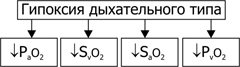
Pic. 25–13 . Changes in gas composition and blood pH during respiratory hypoxia
where P a o 2 and P v o 2 (indicators of oxygen tension in arterial and venous blood), S a o 2 and S v o 2 are indicators of Hb saturation in arterial and venous blood.
¨ Circulatory hypoxia. The reason for the development of cardiovascular (circulatory, hemodynamic) hypoxia is insufficient blood supply to tissues and organs, which is formed on the basis of hypovolemia (decrease in the total blood volume in the vascular bed and heart cavities), heart failure, reduced vascular wall tone, disorders of microcirculation, and oxygen diffusion disorders from capillary blood to cells.
¨ Hemic hypoxia. The reason for the development of blood (hemic) hypoxia: a decrease in the effective oxygen capacity of the blood and, consequently, oxygen-carrying function. The transport of oxygen from the lungs to the tissues is almost entirely carried out with the help of Hb. The greatest amount of oxygen that Hb is capable of carrying is equal to 1.39 ml of gaseous O 2 per g of Hb. The actual transport capacity of Hb is determined by the amount of oxygen associated with Hb and the amount of oxygen given to the tissues. When Hb is saturated with oxygen on average by 96%, the oxygen capacity of arterial blood (V a O 2) reaches about 20% (volume units). In venous blood, this figure is close to 14% (volume units). Consequently, the arterio-venous difference in oxygen is 6%. The hemic type of hypoxia is characterized by a decrease in the ability of Hb red blood cells to bind oxygen (in the capillaries of the lungs), to transport O 2 and to give the optimal amount of O 2 in the tissues. With hemic hypoxia, the real oxygen capacity of the blood can be reduced to 5–10% (by volume).
¨ Tissue hypoxia. Causes of tissue hypoxia: factors that reduce the efficiency of oxygen utilization by tissue cells (usually as a result of inhibiting the activity of biological oxidation enzymes, a significant change in physicochemical parameters in tissues, inhibition of synthesis of biological oxidation enzymes and damage to cell membranes) and / or conjugation of oxidation and phosphorylation.
¨ Substrate type hypoxia. Causes: deficiency in the cells of biological oxidation substrates. In clinical practice, it is often about glucose. At the same time, the delivery of oxygen to the cells is not significantly impaired.
· Resistance of organs to hypoxia. During hypoxia, dysfunctions of organs and tissues are expressed in varying degrees. The tissue of the nervous system has the least resistance to hypoxia.
à Most hypoxia resistant to bones, cartilage, tendons, ligaments. Even in conditions of severe hypoxia, significant morphological deviations are not detected in them.
à In skeletal muscles, changes in the structure of the myofibrils, as well as their contractility, are detected after 100–120 minutes, and in the myocardium, already after 15–20 minutes.
à In the kidneys and liver, morphological abnormalities and disorders of function are usually detected 20–30 minutes after the onset of hypoxia.
à The resistance of nerve cells decreases in the following order: peripheral nerve nodes (for example, in the ganglia of the autonomic nervous system, morphological changes occur in 50–60 minutes) ® spinal cord ® medulla oblongata ® hippocampus ® cerebellum ® cortex of the hemispheres. Termination of oxygenation of the cerebral cortex causes significant structural and functional changes in it already after 2-3 minutes, in the medulla oblongata after 8-12 minutes. It follows that the effects of hypoxia for the organism as a whole are determined by the degree of damage to the neurons of the cerebral cortex and the time of their development.
One of the most important elements of homeostasis of higher animals and humans is oxygen homeostasis. Its essence is the creation and maintenance of an evolutionarily fixed optimal level of oxygen tension in the structures ensuring the release of energy and its utilization.
Oxygen homeostasis is created and maintained by the activity of the system providing the body with oxygen, including external respiration, blood circulation, blood, tissue respiration, and neurohumoral regulatory mechanisms.
Under normal conditions, the efficiency of biological oxidation corresponds to the functional activity of organs and tissues. If this correspondence is violated, a state of energy deficit arises, leading to a variety of disorders, including the death of tissue. Insufficient energy supply of vital processes and forms the basis of a condition called hypoxia.
Hypoxia (oxygen starvation, oxygen deficiency) is a typical pathological process resulting from the insufficiency of biological oxidation and the resulting energy insecurity of life processes. Since a number of organs and systems (respiratory organs, cardiovascular system, blood, etc.) are involved in providing tissues with oxygen, dysfunction of each of these systems can lead to the development of hypoxia. The activities of these systems are regulated and coordinated by the central nervous system, primarily the cerebral cortex. Therefore, violation of the central regulation of these systems also leads to the development of oxygen starvation. Hypoxia is the pathogenetic basis of various pathological conditions and diseases. In any pathological process, hypoxia is present. Since death is a persistent cessation of spontaneous blood circulation and respiration, it means that at the end of any deadly disease, regardless of its causes, acute hypoxia occurs. Dying of the body is always accompanied by total hypoxia with the development of hypoxic necrobiosis and cell death. Oxygen starvation is often the closest cause of the disorder because the oxygen reserves in higher organisms are limited: in humans, approximately 2-2.5 liters. These oxygen reserves, even if fully utilized, are enough for only a few minutes, but dysfunction occurs when there is still a significant content of oxygen in the blood and tissues.
Classification of hypoxia. (Table 1)
Depending on the causes and mechanism of development, the following main types of hypoxia are distinguished.
I. Exogenous hypoxia arising from exposure of external factors to the oxygen supply system - changes in the oxygen content in the inhaled air, changes in the total barometric pressure:
a) hypoxic (hypo-and normobaric),
b) hyperoxic (hyper- and normobaric).
2) respiratory (respiratory);
3) circulatory (cardiovascular) - ischemic and congestive ";
4) hemic (blood): anemic and due to inactivation of hemoglobin;
5) tissue (primary tissue): in violation of the ability of tissues to absorb oxygen, or the dissociation of oxidation and phosphorylation (hypoxia of dissociation).
6). Substrate, (with a shortage of substrates).
7) Overload ("hypoxia load") with increasing load on the oxygen supply system.
8) Mixed.
The flow of hypoxia secrete:
a) lightning (explosive), lasting several tens of seconds, b) acute - tens of minutes, c) subacute - hours, tens of hours, d) chronic - weeks, months, years.
According to the prevalence, there are: a) general hypoxia and b) regional; by severity: a) mild, b) moderate, c) severe, d) critical (fatal) hypoxia.